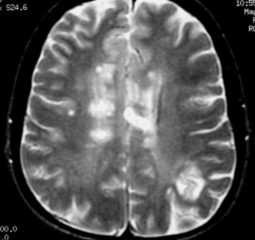
A brain with lesions.
Do you have lesions on your brain? I do not know how many I have. I am not sure but I think my doctor has told me that lesions cause no pain. If that true then why do I constantly have migraines? I think it because my lesions are irritating some nerves.
I was doing some research trying to find out if lesions cause pain. I found some interesting information on lesions. Did you know that that our body try's to re-myelinate? Sometimes it works. It's not back to what it was before but it has some what repaired itself. I have never been told this. It is great to know!
I also learned that I may have "black holes" in my skull! Omg!!!!
I also found out that we should always have contrast when we have an MRI. I have always asked for this when getting an MRI. You will read further done as to why you should have contrast.
MS does its damage by causing the nerves in localized areas in the brain and spinal cord to lose their protective sheaths, called myelin. At first, when the myelin is being attacked, the body brings a higher blood supply to the attacked area or areas to fight the attack, and they becomes swollen and inflamed. These areas now become "lesions." (A "lesion" is simply an area of abnormality in the brain tissue.) If the areas being attacked are large enough they can be seen on an MRI. At this point, when they are inflamed and blood-engorged, they are called "active lesions" because the inflammation is actively attacking the nerve cells. At first the nerves themselves haven't changed much and they appear (and have the same density) as the healthy areas around them. The body attempts to repair the damage that is being done and sometimes these areas try to re-myelinate, with varying success.
They may disappear from the next MRI. They aren't perfect in their function, but the areas may return to a normal appearance.
If the nerves do not re-myelinate and the damage continues, for a long time the lesions sit as scars. These scarred areas contain damaged and dying cells, their blood supply shrinks, and they become dense--more dense than the normal brain around them. These are the classic MS "plaques" and are considered old lesions. They show up as the bright areas, also called "hyperintense," most of us have seen in pictures and on our films. If the attack on the myelin sheath is too strong for the immune system to repair, more and more myelin disappears and the area of nerves eventually dies. Then it completely scars and contracts. The blood flow is decreased to that area and the body tries to reabsorb it. The area then becomes "less dense" than the surrounding normal nerve tissue. After a longer time the scar can reabsorb completely and the area becomes "empty." It's called a black hole. I thought these were just out in space???
Now the very old, mostly dead areas where nerve fibers have died will be seen as less dense (empty) spaces or "black holes". If there are many of these empty areas, the brain will eventually contract and shrink around them. This will be depicted as a loss of brain volume. This is also known as brain atrophy. It is particularly seen in the progressive types of MS. In brain atrophy there will be an increased space between the skull and the brain. The interior area of CSF will be larger. Also the deep folds in the brain (called sulci) will appear widened.
Black holes are areas where a good number of the nerve fibers have been lost. But, not all of them are completely dead. New techniques are showing that black hole can heal and disappear, leaving some scarred tissue behind.
For the most part all visible lesions can be seen without contrast. They show up as T2 Hyperintensities just like all lesions. The problem is that the neurologist can't tell if a lesion is old or new. So new activity will be missed and the MRI may be erroneously dismissed as "unchanged" or "no progression." In the inflammation of new lesions there is a breach in the blood-brain barrier. Wherever there is a break in the blood-brain barrier, the contrast will leak into the brain tissue and the areas will "highlight" or "enhance." They show up as even brighter than the brain around them and brighter than an old, scarred lesion. So new lesions will appear as "enhancing," or "active." Also, older hyperintense lesions that have undergone a new attack at their adges or margins. This is also called reactivation. Lesions that have reactivated may show an even brighter enhancing rim or ring. The appearance of an "enhancing ring or rim" is especially characteristic of MS. When you compare the regular MRI to the contrast MRI you can see the increased brightness of this reactivated, old lesion. New lesions with active inflammation typically show up for 4 to 6 weeks before they scar down and become "old" lesions.
In some cases a newly active MS lesion may not be visible on a regular MRI because the area of nerves, though inflamed, is still pretty much intact and has normal brain density. On the regular MRI it will look like normal brain. Without contrast it won't show up and will likely be missed. When the next phase of MRI is done the contrast is injected into the bloodstream.
There are other techniques such as fast-SPIN, STIR, SPIN-echo, but these are techniques used to clarify tiny differences in the tissue and to make lesions stand out more clearly. That's it in a small nutshell.
I hope you found this information helpful and as knowledgable as I did.
Have a great Sunday! May GOD Bless all of you.
Find me on Facebook!
http://www.facebook.com/christine.thompson
Follow me on Twitter too!
@mywonderMSlife
Thanks for reading my blog!
Christine
 RSS Feed